

Microbiology Society: A world in which the science of #microbiology provides maximum benefit to society | https://teapaw.org/t.co/tMDafCE6df
- Tweets30.5K
- Following1.5K
- Followers76.2K
- Likes12K






ALT Virtual event announcement for Spotlight series on AI in microbiology with guest César de la Fuente and Microbiology Horizons editors on 2 November 2026.

ALT Visual description: A close-up photograph of the face of a whale shark in the ocean. Text reads “Microbe of the Month – Selected from IJSEM” and “Ureaplasma rhincodontis”. A caption below explains that it is a novel bacterial species isolated from whale sharks that is the first of its kind found in "cold-blooded" vertebrates.

ALT Fig. 2. Ziehl–Neelsen staining – microscopic observation at 100× magnification (immersion) of AFB.

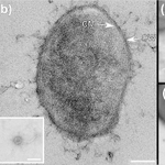
ALT Fig. 3. Phase-contrast and electron micrographs of Methanomonile shimae (a, b), Methanobrevibacter ruminantium (c), Methanocatella smithii (d), Methanobaculum cuticulare (e) and Methanobinarius arboriphilus (f). The inset in the ultrathin section (b) shows a virus from a negative stain of the same culture. Also the boxed regions in c–f (negative stains) indicate virus particles. CM, cytoplasmic membrane; CW, cell wall. Scale bars: 5 µm (a), 0.5 µm (b–f), 0.1 µm (insets).

ALT Virtual event announcement for Spotlight series on AI in microbiology with guest César de la Fuente and Microbiology Horizons editors on 2 November 2026.


ALT Fig. 1. Antimicrobial susceptibility testing results of 96 strains of K. pneumoniae.

ALT Fig. 2. Genome organization of selected bacilladnaviruses. ORF colours: Rep, cyan; CP, green; putative phospholipase, pink; hypothetical proteins, grey.

ALT Fig. 2. ML phylogenetic tree of ST3 A. baumannii isolates. The tree is generated from recombination-filtered core-genome SNPs. Metadata, including country, year of isolation, source, KL and OCL types, ampC alleles and resistance genes, is shown on different rings around the tree. ‘NK’ indicates ‘not known’. Distinct clades and subclades are shaded in different colours, with subclade designations indicated inside the shaded areas. Colour coded key is shown.

ALT Microbiology Society and New England Biolabs logos promoting Vacation Studentships Grant Partners program.

ALT Covid-19 Restrictions Dublin stock photo. Credit: iStock/lensmen


ALT Microbiology Society's 2027 Annual Conference welcome banner with abstract submission deadline on 10 November 2026.


ALT Virtual event announcement for Spotlight series on AI in microbiology with guest César de la Fuente and Microbiology Horizons editors on 2 November 2026.

ALT Fig. 1. Geographic sampling and host-virus co-phylogeny. (a) Map of collection sites. Detailed coordinates of each location are provided in Methods. (b) Stacked bar charts of PCR-positive samples by location and sample type. Each facet corresponds to a specific site, with colours indicating bat species. (c) Tanglegram of co-phylogeny.

ALT Banner for the Microbiology Society Annual Conference 2027 with abstract submission deadline on 10 November 2026, inviting attendees to Birmingham.