
A pediatric radiology textbook, one case every day. Opinions are my own. #FOAMed #FOAMPed #FOAMRad #MedEd #PedsRad #radiology #radiología #radiologie
Joined September 2014
- Tweets8.3K
- Following23
- Followers13K
- Likes0






Pinned Tweet
#FOAMed #FOAMPed #FOAMRad #MedEd #PedsRad #RadEd #RadRes #radiology #radiología #radiologie #pediatrics #paediatrics #pediatría #pédiatrie #pädiatrie
We are publishing a pediatric imaging textbook, one case every day, and currently have 2,000 cases covering 1,000 diseases and 125 differential diagnoses, 50 procedure descriptions and 4 review courses.
You can find us on the Web (pediatricimaging.org) and to see every case follow us via RSS (pediatricimaging.org/feed) or via email (enter your address + click "Follow" button at the bottom of the page at pediatricimaging.org).

School ager with palpable mass behind knee
Transverse grayscale (above)+sagittal color doppler (below) US of popliteal fossa show large anechoic lesion in popliteal fossa...
#FOAMed #MedEd #FOAMRad #PedsRad #RadEd #radiology #pediatrics #Ultrasound #Orthopedics #Orthopaedics

...which is avascular and whose neck at its deepest extent was seen to extend into the joint space between the semimembranosus tendon and medial head of gastrocnemius tendon.
The diagnosis was popliteal cyst.
Learn more: pediatricimaging.org/disease…
In need of a review of pediatric fractures?
Clicking on Pediatric Fractures+Dislocations will take you to descriptions of approaches to+appearances of > 65 #pediatrics fractures+dislocations
Try it at PediatricImaging.org/fractur…
#FOAMRad #radiology #EmergencyMedicine #Orthopedics

Newborn with posterior fossa cyst on prenatal US
Sagittal T1 MRI(above left) shows cystic lesion in inferior posterior fossa which is not causing any mass effect in normally sized posterior fossa
#FOAMed #FOAMRad #PedsRad #RadEd #radiology #pediatrics #Neurology #Neurosurgery

Coronal T2 MRI (above right) and axial T1 MRI without contrast (below) show a mildly enlarged fourth ventricle which is connecting to the cystic lesion due to mild agenesis of the cerebellar vermis.
The diagnosis was Dandy Walker variant.
Learn more: pediatricimaging.org/disease…
Preschooler with elevated liver function tests
Sagittal (above left)+transverse (above right) US of liver show increased echogenicity around portal veins - a starry sky appearance
#FOAMRad #PedsRad #RadEd #radiology #pediatrics #radiologia #PedSurg #SoMe4PedSurg #Ultrasound

Sagittal (below left) and transverse (below right) US of the gallbladder show diffuse gallbladder thickening.
The diagnosis was acute hepatitis.
Learn more: pediatricimaging.org/disease…
Newborn who experienced shoulder dystocia during delivery
CXR AP shows a complete fracture through the diaphysis of the left humerus.
#FOAMed #MedEd #FOAMRad #PedsRad #RadEd #radiology #pediatrics #radiologia #NICU #Neonatology #Ortho #Orthopedics #paediatrics #pediatria

The fracture fragments are not in anatomic alignment and apposition.
The diagnosis was proximal humerus fracture.
Learn more: pediatricimaging.org/disease…

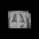
Toddler with respiratory distress
CXR AP (left)+CXR lateral (right) show heart to be enlarged+pulmonary vascularity to be increased.
#FOAMed #MedEd #FOAMRad #PedsRad #RadEd #radiology #pediatrics #radiologia #medstudent #medicalstudent #radiologie #paediatrics #pediatria

The lungs are clear.
The diagnosis was atrial septal defect.
Learn more: pediatricimaging.org/disease…

Newborn with respiratory distress
CXR AP is angled superiorly at greater than 90 degrees to the patient and therefore shows the apices of the lungs very clearly.
#FOAMed #FOAMRad #PedsRad #RadEd #radiology #pediatrics #radiologia #NICU #Neonatology #paediatrics #pediatria

The ribs have a different contour than would be expected on a normal CXR AP angled at 90 degrees to patient. Heart size is normal, pulmonary vascularity is not congested+lungs are clear.
The diagnosis was normal lordotic view of chest.
Learn more: pediatricimaging.org/disease…
Starting a pediatric radiology rotation, preparing to take call, or in need of a neonatal imaging review?
Try it at PediatricImaging.org/neonata…
#FOAMed #MedEd #FOAMRad #PedsRad #RadEd #RadRes #radiology #radiologia #pediatrics #paediatrics #pediatria

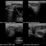
Newborn with bilateral unpalpable testicles
Transverse(above left)+sagittal(below left) US of pelvis show right testicle to lie lateral to superior aspect of bladder
#FOAMed #MedEd #FOAMRad #PedsRad #radiology #pediatrics #radiologia #NICU #Neonatology #Ultrasound #paediatrics

Transverse (above right) and sagittal (below right) US of the left inguinal canal shows the left testicle to lie within the left inguinal canal.
The diagnosis was bilateral undescended testicles with the left testicle in the inguinal canal and the right testicle in the abdomen.
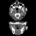
School ager with left preauricular mass
Axial (above)+coronal (below) CT show well-circumscribed oval lesion near left ear that has calcifications within it.
#FOAMed #MedEd #FOAMRad #PedsRad #RadEd #radiology #PedSurg #SoMe4PedSurg #pediatrics #radiologia #paediatrics

There are no inflammatory changes associated with it.
The diagnosis was pilomatricoma.
Learn more: pediatricimaging.org/disease…
