

BJJ, edited by Professor Fares Haddad, is the flagship journal published by The British Editorial Society of Bone & Joint Surgery, a registered charity.
- Tweets14.1K
- Following1.5K
- Followers35.8K
- Likes2.7K




ALT Webinar series by The Orthopaedic Trauma Society on trauma care, featuring live sessions every other Wednesday starting January 6, 2027.

ALT Serial anteroposterior radiographs of an 18-month-old male with initially non-fractured Crawford type III congenital pseudarthrosis of the tibia. a) An initial radiograph at presentation shows an intact cortex without a radiolucent line. b) A radiolucent line (arrow) was evident at the three-month follow-up. c) It progressed to a cortical fracture (arrow) at the 6.75-month follow-up. d) Extension of the fracture (arrow) at subsequent follow-up.

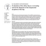
ALT Abstract text for article titled 'Evaluation of the clicky hip as a screening tool in the diagnosis of developmental dysplasia of the hip'. The Bone & Joint Journal

ALT Webinar series on limb reconstruction management techniques with next session on Tuesday 27th October, offering 31 CPD points from the Royal College of Surgeons of England.

ALT BJJ Paper of the Month highlighting venous thromboembolism risk after knee, hip, or shoulder arthroplasty by Swain et al.

ALT Raikin nine-zone system for localization of osteochondral lesions of the talus.

ALT The European Bone & Joint Infection Society recommendations for debridement, antibiotics, and implant retention (DAIR) in periprosthetic joint infection (PJI).11 The three-level recommendation allocates patients to either the green group, with good indications for DAIR, the orange group with indications which, if not addressed, may carry a higher risk of failure, and the red group, where DAIR would not be recommended.

ALT Radiostereometric analysis measurement for medial and lateral minimal joint space width (mJSW) and the coordinate system for the migration measurements of a right tibial and femoral component.

ALT Femoral registration of robotic unicompartmental knee arthroplasty to total knee arthroplasty.

ALT BJJ Life logo with a green leaf and tagline about orthopaedic MCQ resources from The Bone & Joint Journal above a Quickstart button.

ALT Abstract text for article titled 'The impact of changing Coronal Plane Alignment of the Knee classification on early functional outcomes after primary robotic-assisted total knee arthroplasty'. The Bone & Joint Journal


ALT Abstract text for article titled 'Higher femoral periprosthetic fracture rate with dual-mobility compared with single-mobility total hip arthroplasty'. The Bone & Joint Journal

ALT Close-up of a professional microphone with The Bone & Joint Journal Podcasts logo and icons for Apple Podcasts, Spotify, YouTube, and Amazon Music below.

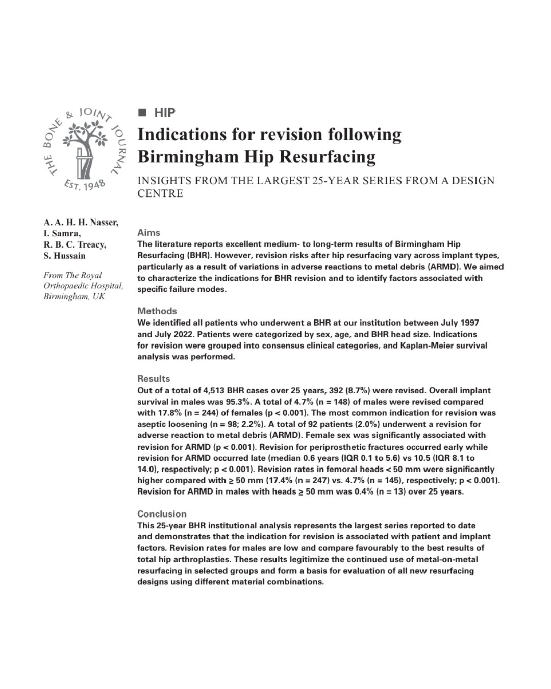
ALT Abstract text for article titled 'Indications for revision following Birmingham Hip Resurfacing'. The Bone & Joint Journal

ALT Dynamic radiological images (top row) were obtained using a flat panel X-ray detector to capture the motion of the hip during racket swinging. The corresponding 3D models (middle row) were aligned to each radiological image using a model-image registration technique, allowing visualization of the in vivo relative motion between the pelvis (blue) and femur (orange).

ALT European Hip Society's live debate on young adult hip surgery: preserve or replace earlier, moderated by Professor Carsten Perka.