

Eye MD | Fixing Eyes in Kashmir ❤️ | Cataract & Retina
Jammu And Kashmir
Joined March 2020
- Tweets1.3K
- Following213
- Followers1.1K
- Likes1.8K


Pinned Tweet
𝐑𝐞𝐭𝐢𝐧𝐚𝐥 𝐃𝐞𝐭𝐚𝐜𝐡𝐦𝐞𝐧𝐭
Hope you guys can appreciate the detached retina and also a retinal hole.
Smartphone video capture simplifies patient education
#Ophthalmology #Retina #Retinaldetachment #MedTwitter #MedEd
What can a 4 sec clip tell you about this eye ??
Share your thoughts
#Ophthalmology #EyeDoc #Eyes
#EyeEd
Extreme Capsulophimosis
Although there are several reasons for capsulophimosis, such extreme cases are rare. What would you speculate was the cause here ??
#Ophthalmology #Cataract #EyeEdu #Eyedoctor


3 Seconds to Diagnose this 👇🏻
What’s your diagnosis #Ophthalmologists ??
#eyecare #eyes #Ophthalmology
Peaked Pupil -Post Sx 🚩
🔹 Iris incarceration/prolapse
🔹 Vitreous prolapse /PCR
🔹 Iridodialysis
🔹 Sphincter tear
🔹 IOL-related iris capture
🔹 Retained lens matter/inflammation
🔹 Early posterior synechiae
#CataractSurgery #Ophthalmology #PostOpComplications

“A year of learning, sharing and growth. Thankful for every mentor and every platform”
Happy New Year to All
Capsulophimosis:
• Small CCC (<5 mm) biggest risk factor.
• Watch early for capsular edge fibrosis/whitening.
• YAG radial relaxing cuts (4–6) first-line.
• Dense fibrosis → surgical membranectomy.
• Prevention: 5–5.5 mm CCC, CTR in PXF/zonular weakness.


Myopic Foveoschisis
1️⃣ Splitting of retinal layers in high myopia
2️⃣ OCT = gold standard diagnosis
3️⃣ Symptoms: blur, metamorphopsia (may be silent early)
4️⃣ Risk: macular hole / RD progression
5️⃣ Tx: PPV ± ILM peel, gas; observe if stable
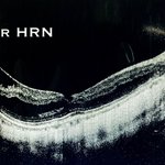
This is a very interesting OCT Scan.
What all can you tell me about it ??
What’s happening here ?? What to do about it ??
#Ophthalmology #MedTwitter #Retina
#MedEd

This is a very interesting OCT Scan.
What all can you tell me about it ??
What’s happening here ?? What to do about it ??
#Ophthalmology #MedTwitter #Retina
#MedEd
Acute anterior uveitis + hypopyon → think of:
1️⃣ HLA-B27 uveitis
2️⃣ Behcet’s disease (shifting sterile hypopyon)
3️⃣ Infective endophthalmitis (purulent)
4️⃣ Tuberculosis / Sarcoid (granulomatous)
5️⃣ Herpetic uveitis (with keratitis/↑IOP)
#Ophthalmology #MedTwitter


"1 in 20 people with diabetes may lose clear vision to Diabetic Macular Edema. Don’t let sugar steal your sight — regular eye check-ups can protect your vision."
#SugarVsSight
#WorldRetinaDay

𝐀𝐧𝐭𝐞𝐫𝐢𝐨𝐫 𝐂𝐡𝐚𝐦𝐛𝐞𝐫 𝐈𝐎𝐋
Checklist is you see this patient ▶️
1️⃣ Watch corneal endothelium (edema / cell loss
2️⃣ Check IOL position & stability
3️⃣ Examine angle (PAS / crowding / glaucoma)
4️⃣ Look for iris changes (pigment / UGH)
5️⃣ Monitor IOP & optic nerve

Diagnosis :
• Epiretinal Membrane
• PseudoMacular Hole
• Splitting of Retinal layers
Management :
• Mostly observe unless significant metamorphosis/DOV
• In Severe Cases : VIT+ERM Removal
💎 Observe till you can
#Ophthalmology #MedTwitter

👁️🗨️ Granular Corneal Dystrophy
🍞 “Breadcrumb” white stromal deposits
✨ Clear spaces between opacities
🧒 Onset 1st–2nd decade, slow progression
🚫 Recurrent erosions rare (vs lattice)
🔬 PTK early | DALK/PKP if advanced


Dendritic Keratitis ❄️
1️⃣ Branching epithelial ulcer with terminal bulbs = classic sign
2️⃣ Steroids worsen → geographic ulcer
3️⃣ Reduced corneal sensation
4️⃣ Treat with antivirals, not antibiotics
5️⃣ Recurs often → consider oral prophylaxis
#Ophthalmology #medtwitter
