

#Vitreoretinal Surgeon | @uiowaeye, Cinci Eye, @umiamimedicine @FIUbiomed alum | Chronicling what I learn along the journey (but tweets =/= medical advice)
Miami, FL
Joined April 2016
- Tweets485
- Following45
- Followers660
- Likes68





I always screen patients for migraine especially after a negative cardiovascular work up. Perhaps a vasospasm-mediated mechanism. Migraine w/aura higher risk than w/o aura.

This is the first large study to show that migraine is associated with increased risk of a central, branch, and other types of retinal artery occlusion (RAO) after adjusting for all known risk factors of RAO.
sciencedirect.com/science/ar…
#retina #migraine #cardiology #neurology
This, along with reduction in chance of retinal folds, is why I seldom recommend strict face down positioning following vitrectomy.

MRI imaging demonstrated that side positioning gives better contact between the gas bubble and the inferior and anterior retina than prone positioning even when the gas fill is only 70% of the vitreous cavity. N=3, MRI performed on post op day 1 ow.ly/4hH150zko7O

Tips for sutureless intrascleral haptic fixation from Ashkan Abbey for the VR surgeon:
1) thorough PPV w/base shave @ sclerotomy sites (prevent vit incarceration when pulling haptic out)
2) inject IOL into vit cavity
3) MaxGrip forcep - grab tip of haptic in PARALLEL fashion
4) pull trochar cannula out BEFORE externalizing haptic
5) create flange (⬇️ risk dislocation) & tuck in
6) create iridotomy (⬇️ chance of reverse pupillary block)
7) second haptic is hardest to secure; if trouble viewing in vit cavity, push on the optic
#VBS2019 @RetinaToday
Efren Mandelcorn presents a rapid way of closing leaking sclerotomies with a 30ga needle puncture through the offending sclerotomy ncbi.nlm.nih.gov/m/pubmed/30… @VitBuckle #VBS2019
Judy Chen shows a slick PFO-less technique to repair an inferior PVR RD using “struts”: create 2 rows of diathermy 180-200 degrees and use soft tip to lyse retina but leave 2-3 areas attached before FAX to avoid slip, then drain through the “struts” #VBS2019

Great tips, along with a paper discussing a role of dilute intravitreal povidone-iodine ncbi.nlm.nih.gov/pmc/article… @VitBuckle #VBS2019


Dr. Shriji N. Patel outlines the keys for success in vitrectomy for endophthalmitis at #VBS2019. He says you should plan plan plan, that view is everything, infusion is your lifeline, and work front to back.

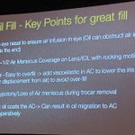
Great tips for maximizing SO fill via Emmanuel Chang - use the nasal trochar to inject oil, tilt the eye nasal and vent temporally @VitBuckle #VBS2019

Pravin Dugel recommends using an agent with the HIGHEST surface tension (air, has) to tanponade a large intraoperative retinal bleed after raising IOP and coagulation have failed (or not an option) #VBS2019
Tom Albini of @BascomPalmerEye stresses that oral steroids need to be tapered to 10mg/daily after TWO (max 3) months when treating uveitis; if recurrence occurs, a second immunomodulator is required #RetinaFellowsForum2019
Dean Eliott of @MassEyeAndEar gives us some nuances between epiretinal membranes vs proliferation #RetinaFellowsForum2019

When using the fragmatome to remove retained lens fragments, John Kitchens recommends first LOWERING the power to reduce chatter #protip #RetinaFellowsForum2019
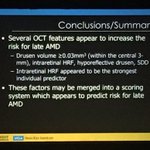
Carl Awh discusses the nuances of genetic testing in AMD. Key points:
1. Macular genetic panels for AMD only matter IF the patient has a diagnosis of AMD
2. High CFH, low ARMS2 risk alleles levels portend a ⬆️ progression to neovascular AMD in patients taking AREDS formulation
Tom Albini gives us the key signs for primary intraocular lymphoma. Think about it when you see a lot of larger vitreous cells without much other uveitis symptoms or signs (i.e., white, quiet eye) #RetinaFellowsForum2019

Albini recommends using a lab that is used to dealing with OCULAR samples versus a general pathology lab #RetinaFellowsForum2019
