



Quique Revilla retweeted

Do you think making a diagnosis of myeloid/lymphoid neoplasms with tyrosine kinase fusions (MLN/TK) is challenging?🤯 Join the club... so do we 😉. So we decided to do something about it and create a user-friendly guide! Out today in @AJCPjournal with
@jenweiying we hope you find this helpful. Make sure to check out the supplemental file. It has a useful cheat sheet. Free access link shorturl.at/rwBuB

Quique Revilla retweeted

Even without a good size reference, this small aggregate of cells is adequate for the diagnosis of a metastatic adenocarcinoma from the patient's known lung primary. (CSF)

Quique Revilla retweeted

64 yo male with abdominal pain and ascites. On CT - infiltration of the fat around the aorta and the perinephric fat and omental cake. This is a biopsy from the perinephric fat. Thoughts? – at Israel


Quique Revilla retweeted

WHO 2026 Clarification on Immature Teratomas
Any amount of immature neuroepithelial tissue in a teratoma → diagnose as immature teratoma
Before calling it immature, rule out mimics:
- Cerebellar tissue (pic 1)
- Retinal tissue (pics 2 & 3; pic 3 was previously misdiagnosed as immature teratoma)
- Ependymal tissue (pic 4)
Dr. McCluggage. 2026 Annual Meeting of the International Society of Gynecological Pathologists
#ISGyP #Pathology #PathX


Quique Revilla retweeted

What I'm learning at #SHEAHP2019
When assessing a MPN-like morphology with increased eos on marrows, also look at mast cells. Abnormal mast cell spindled morphology or phenotype may clue you into an underlyng PDGFRb rearrangment MLN. Esp. if KIT mutation absent.
#hemepath


Quique Revilla retweeted

#BreastCarcinoma being gobbled up by lymphocytes. #InvasiveBreastCarcinomaNSTWithMedullaryPattern
This one also shows medullary pattern #DCIS 📸3
Hope y’all having a great weekend!
💙
#pathology




Quique Revilla retweeted

60 y/o with PMHx of obesity
10/10 pain in MTP joint of big toe
Synovial fluid below with 30,000/uL WBCs👇
Dx? 🤔 #arthritis #gout #pseudogout #usmle #chemistry #pathagonia #synovial


Quique Revilla retweeted

Series 4 : Diagnostic challenges in Plasma Cell Neoplasms .
📌Post 2 : Morphology Pitfalls in Plasma Cell Neoplasms- 2026 review.
📌Most myelomas are easy. The variants are not.
⏩Small cell / lymphoplasmacytic variant
- 3-4% of cases
- Looks like LPL or MZL
- Frequently CD20+ and cyclin D1+ ( because of t(11;14))
- Key separators : CD138+ , usually CD45- , CD19-, clinical CRAB / lytic lesions , no MYD88.
⏩Plasmablastic variant
- Large vesicular nuclei , prominent nucleoli
- almost identical morphology & phenotype to Plasmablastic lymphoma .
- Differentiate by clinical context , extent of marrow involvement , M- protein , FLC ratio and FISH ( gain 1q , del 1p , t(4;14) favour myeloma .)
⏩Anasplastic / pleomorphic variant
- CaN express CD30 and mimic ALCL
- Rare aberrant T cells markers ( CD3, CD4 ) occur.
⏩Myelomatous Effusion
- can occur at initial diagnosis or during progression ( an ominous sign ).
- Even when plasma cells become multinucleated or pleomorphic , the classic eccentric nucleus and perinuclear hof often persist.
- When Morphology turns Plasmablastic , always Differentiate from primary effusion lymphoma , Plasmablastic lymphoma and fluid overload large B cell lymphoma .
- Presence of M- protein + CRAB features strongly favours Myelomatous Effusion .
⏩Plasma Cells Leukemia
- Defined by > 5% circulating plasma cells in peripheral blood ( WHO - HAEM5 / IMWG 2021 )
- Primary PCL ( de novo ) is highly aggressive.
- Secondary PCL develops during relapse or progression .
- Leukemic plasma cells are frequently CD56-negative.
- Emerging data suggest even > 2% circulating neoplastic plasma cells may behave like occult primary PCL .
Save this Post for quick reference
#Hematopathology #Myeloma #Pathology #PlasmaCellLeukemia #DiagnosticPathology #MultipleMyeloma #Morphology


Quique Revilla retweeted

Esta semana nos ha tocado despedir a la Dra. María Cámara (@mcamarajurado). Después de 14 años con nosotros, te damos las gracias por tu dedicación, tu compañerismo y tu contribución a nuestro servicio. Te deseamos lo mejor en esta nueva etapa. ¡Esta siempre será tu casa!

Quique Revilla retweeted

Extra-nodal Rosai Dorfman disease shows overlapping features with IgG4-related fibrosclerosing disease - storiform fibrosis and inflammation. The S100 stain highlights emperipolesis with nuclear (short arrows) and cytoplasmic staining; engulfed cells are negative (long arrows).


Quique Revilla retweeted

Cytomegalovirus on ROSE. The inclusions are evident on DQ and Pap. Cell block is loaded. CMV stain is great too! 👀🦉🔬 #cytology #cytorocks

Quique Revilla retweeted

ALCL in a pericardial effusion🤩CD30, CD3, CD4, CD5, CD45+. Gorgeous! #hematology #hematopathology #pathology #oncology #alcl #hemepath @NathanPaulsonMD @AadilAhmedMD @MaracajaDLV


Quique Revilla retweeted
“In CSF, size matters the most, especially when the nuclei look guilty.” Metastatic breast lobular carcinoma. (cytospin)

30 yo woman with a complete hydatidiform mole one month ago. Endometrial aspiration revealed retained placental tissue. Is this atypical trophoblastic proliferation sufficient to diagnose choriocarcinoma? Opinions, please. #gynpath


Quique Revilla retweeted

Remember that Acute erythroid leukemia can be CD34 positive, don’t exclude a diagnosis of AEL/PEL because you see CD34, by Dr Wei Wang


Quique Revilla retweeted

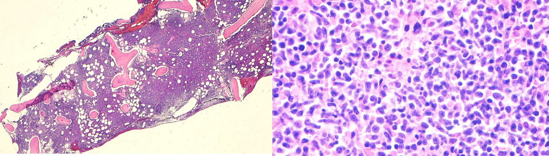
Mantle cell lymphoma in a bone marrow biopsy. I love it when things work out well!
#pathology #hemepath👩🔬🩸


Quique Revilla retweeted

T-LGL leukemia. STAT3 p.Y640F +. Nice linear arrays of granular lymphocytes expressing CD8, TIA, and granzymeB. This is a nice @BloodJournal paper from @moricemdphd describing these findings (https://teapaw.org/t.co/RGStmDvgwS). #hemepath @SocforHemepath @hematogones


