

MBBS,MD,DNB(Radiology)|Fellowship in MSK Imaging|Interested in AI ML|Cricket|Football| Finance| Fitness| Photography| ISTP-T |Grateful🙏
India
Joined November 2020
- Tweets836
- Following589
- Followers242
- Likes5.9K





Pranav Udupi retweeted

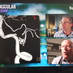
1/Talk about twisting your back!
Do spine vascular lesions make your brain feel as tangled as the dilated vessels you see?
Want some information on malformations?
Here’s a thread on spine vascular anatomy to give you durable knowledge on dural arteriovenous fistulas (dAVF)


Pranav Udupi retweeted

#ASHNRCOTW 320: ANSWER. Thx Meltzer 4 case! #ASHNR26
@callyrobs @DShatzkes @CDP_Rad @rhwiggins
@KRileyMD @CMGlastonbury @tabby_kennedy
@PhilipRChapman1 @cmtomblinson @amyfjuliano
@bpoliceni @MohitTCCNeuro @AshokSrini15 @KatieTraylorDO @CynXinWu


Pranav Udupi retweeted

👨🏫 Let us learn today about a not so oft talked | recognized cause of acute ischemic stroke.
We should always ⚠️: the vessels, their sources & their constituents are the mischief mongers.
🧠 is just their ⚽️ 🏟️
57♀ H/o allergic asthma, COVID pneumonia.
1 day h/o vision disturbance, confusional state.
MR 👇
1|14

Pranav Udupi retweeted

#NeuroTips
Venous developmental anomalies (DVAs) can go south...
As in this case of left temporal hemorrhage due to thrombosis of the collecting system of a DVA.
Be careful with stroke in young patients!
📦Think outside the box!

Pranav Udupi retweeted

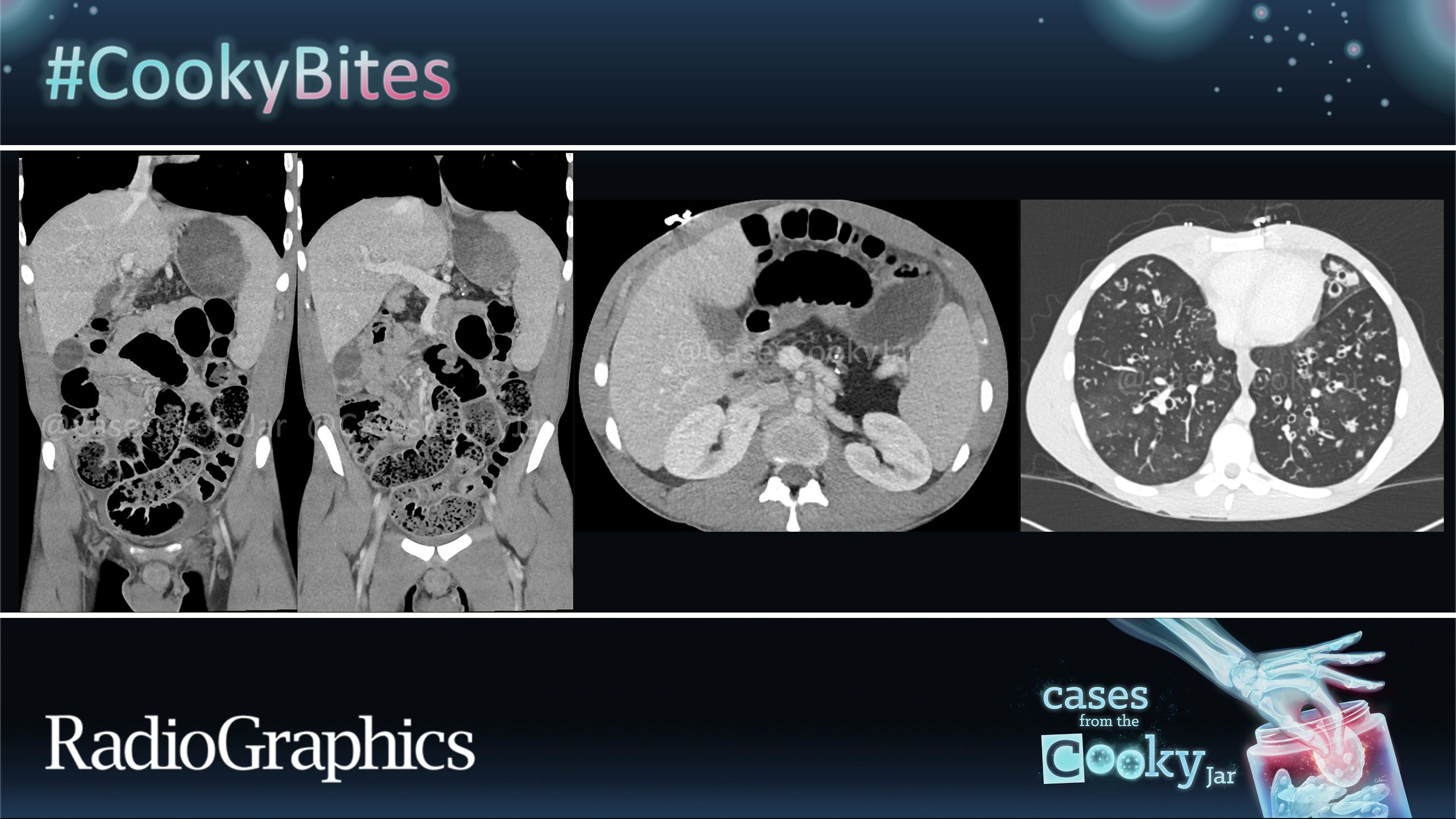
22 YO male presents with recurrent bouts of abdominal pain.
What is the diagnosis?
We’ll post the answer in 24h. Share companion cases with us using #CookyBites #239. We will RT the best cases!
#RGphx @cookyscan1 @RadioGraphics
Pranav Udupi retweeted

🎯 Not all calf tears are classic.
Here are three unusual calf injuries on ultrasound - each with its own diagnostic challenge.
#MSKUltrasound #SportsUltrasound #SonographerEducation #RadiologyEducation #CalfInjury #UltrasoundCase #sonography #radiography #sonographer
Pranav Udupi retweeted
We have all seen a carotid (fetal) origin of the posterior cerebral artery...
But what about a basilar origin of the middle cerebral artery over a homolateral carotid agenesis?
Nature never ceases to amaze us.
#NeuroTips


Pranav Udupi retweeted

Here is the summary of a clinicoradiologic approach to diagnosing the etiology of myelopathy

Pranav Udupi retweeted

Awesome review of Pulmonary Aspiration Syndromes in the July issue of JTI. Here's a summary. Check out the full article for details. tinyurl.com/38fwzdvr @ThoracicImaging


Pranav Udupi retweeted
#ASHNRCOTW 314: ANSWER. Thx Drs. Warren + Vaysberg 4 case! #ASHNR25
@callyrobs @DShatzkes @CDP_Rad @rhwiggins
@nakoontz @KRileyMD @CMGlastonbury @tabby_kennedy @PhilipRChapman1 @cmtomblinson
@amyfjuliano @bpoliceni @MohitTCCNeuro @AshokSrini15 @KatieTraylorDO @CynXinWu
@WmOBrienDO @RadRupa


Pranav Udupi retweeted

#TCCCoffeetimevid Neck spaces parch your throat? Here is a short and simple tutorial on neck spaces to make it quick and easy. Enjoy it at coffee time! Take the short quiz at the end of the video to test your knowledge.
@thecortexclub @RamVaidhyanath @MCWNeurorad @ASHNRSociety @ESHNRSociety @teamishnr @RANZCRcollege #radres @RadDiscord
Pranav Udupi retweeted

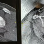
The Six Types of Bankart and Bankart-Variable Lesions
a) Complete detachment of the antero-inferior labro-ligamentous complex
b) Bony Bankart (avulsion of osseous fragment from the antero-inferior glenoid)
c) PERTHES lesion
d) ALPSA lesion
e) GLAD lesion
f) HAGL lesion

Pranav Udupi retweeted
A weightlifter developed intense back and abdominal pain during competition.
An ultrasound led to CT urography. Have you even heard of this diagnosis before?
Images in 𝑅𝑎𝑑𝑖𝑜𝑙𝑜𝑔𝑦

Pranav Udupi retweeted
How durable is your knowledge of dural lesions?
How’s your knowledge base of the skull base?
Know the difference between petroclival & petrous face? Middle fossa & middle sphenoid wing? Clinoid & clivus?
Here is a map of the names of skull base meningiomas. How many do you know?
Recognizing a lesion is only the first step. Communication is the next step.
If you can’t accurately communicate where a lesion is located, then you have only done have your job.
When it comes to skull base meningiomas, high risk lesions are the medial lesions:
Clinoid
Cavernous sinus
Tuberculum sellas
Medial/incisural tentorium
Clivus
I think of a cone coming up from the foramen magnum encompassing these lesions that I call the “cone of risk”!
Hopefully, this anatomy of skull base dural lesions with stay with you for the duration!

Pranav Udupi retweeted

Teaching NeuroImage: Subacute Encephalopathy With Seizures in Alcoholics (SESA)–Related Abnormalities on EEG and MRI | Neurology neurology.org/doi/10.1212/WN…

Pranav Udupi retweeted

"“Flow Void Sign”: Flow Artifact on T2-Weighted MRI Can Be an Indicator of Dural Defect Location in Ventral Type 1 Spinal CSF Leaks"
doi.org/10.3174/ajnr.A8445
@NeuroradLal #CSFLeak #FellowsJournalClub


Pranav Udupi retweeted

Happiness is highly linked to how focused you are on what you're doing.
A wandering mind is an unhappy mind.

