

Your Guide to Health And Wellness. A podcast Every Week. Check out our podcast: https://teapaw.org/t.co/nplGVFMDMw
Africa| Contact+250789848972
Joined June 2021
- Tweets3.9K
- Following50
- Followers3.9K
- Likes65K



RWANDA & UNICEF: 40 YEARS OF PARTNERSHIP IN MATERNAL AND CHILD HEALTH
Rwanda’s Minister of Health, Dr. Sabin Nsanzimana, has received Etleva Kadilli, UNICEF Regional Director for Eastern and Southern Africa, for discussions on strengthening cooperation in healthcare and human capital development.
The meeting comes as UNICEF marks 40 years of partnership with Rwanda and 80 years globally.
Discussions highlighted UNICEF’s longstanding and ongoing contribution to maternal and child health, alongside efforts to strengthen human capital.
The partnership raises an important question for Rwanda’s health sector: How can decades of cooperation translate into better maternal health outcomes, improved child survival, stronger nutrition services and equitable access to quality healthcare?
#HealthNews #Rwanda #UNICEF #MaternalHealth #ChildHealth

65
Yesterday was Cervical Cancer Elimination Day.
Cervical cancer is largely preventable, yet it continues to claim lives across Africa.
Africa CDC calls for accelerated action to protect girls through HPV vaccination and ensure women have access to screening, timely treatment and care
Together, we can eliminate cervical cancer.
#EndCervicalCancer

82
Girls In Rwanda Are Still Dropping Out of School Due to Lack of Sanitary Towels!
youtu.be/IkqlF8XT6tI?si=c3pN…
125
THE CLITORIS IS MUCH MORE THAN THE SMALL PART YOU CAN SEE.
Many people think the clitoris is just a tiny button located above the vaginal opening. But its anatomy extends much further inside the body.
Here is what you should know:
1. The glans: This is the visible external part of the clitoris. It contains thousands of sensory nerve endings and is highly sensitive.
2. The body (shaft):** Located beneath the surface, it connects the visible glans to the deeper structures.
3. The crura: These are two internal extensions that branch out on either side, following the pelvic bones.
4. The vestibular bulbs: These are paired erectile tissues located on either side of the vaginal opening. They are closely associated with the clitoris and become engorged with blood during sexual arousal.
WHAT DOES THE CLITORIS DO?
Its primary known function is sexual sensation and pleasure. During sexual arousal, its erectile tissues become engorged with blood, just like other erectile tissues in the body.
AN IMPORTANT FACT:
The clitoris is not simply a small external organ. It is a complex anatomical structure, much of which is hidden from view.
Understanding clitoral anatomy helps promote accurate sexual health education, informed conversations about the body and better understanding of female sexual health.
Knowledge of the human body should never be a taboo.
#HealthEducativeContent #SexualHealth #Anatomy #WomensHealth #Rwanda
379
To save lives and make #CervicalCancer history, we call on all countries to ensure that:
Every girl can get an HPV vaccine.
Every woman knows when she is due to be screened.
Every woman diagnosed receives the care she needs without delay.
189
Sad News:
More than 11,400 Ebola cases have been reported in #DRCongo, and nearly half of those infected have died.
Despite progress in the response, the outbreak continues to spread.
Here are 5 vital parts of the efforts to stop Ebola. 👇
172
WHAT HAPPENS WHEN YOU DRINK PROTEIN ON AN EMPTY STOMACH?
Drinking a protein shake before eating anything else can have some interesting effects on your body.
Here are 5 things to understand:
1. Muscle recovery: Protein provides amino acids that help repair and build muscles, especially after exercise.
2. Keeps you fuller: Protein stimulates satiety hormones, helping you feel satisfied and potentially reducing hunger.
3. Digestion and absorption: Your body can digest and absorb protein without requiring you to eat other foods first. An empty stomach does not automatically make protein more effective.
4. Blood sugar regulation: Protein can slow gastric emptying and influence blood sugar responses, particularly when consumed alongside carbohydrates.
5. Not necessarily better for weight loss: Drinking protein on an empty stomach does not directly burn more fat. Your overall calorie intake and physical activity matter more.
IMPORTANT: Protein shakes are not a substitute for a balanced diet. Whole foods such as eggs, beans, fish, milk and nuts also provide valuable nutrients.
THE TAKEAWAY: You don't need to eat before drinking protein. What matters most is your total daily protein intake, the quality of your diet and your physical activity.
Your body doesn't care whether you drink protein first thing in the morning or after breakfast. It cares about getting the nutrients it needs.
#HealthEducation #Nutrition #Protein
4
343
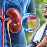
YOUR KIDNEYS CAN BE FAILING WITHOUT YOU FEELING ANY PAIN.
One of the dangers of chronic kidney disease is that it can develop gradually, sometimes without noticeable symptoms until the condition becomes advanced.
No pain does not automatically mean healthy kidneys.
Signs that may suggest your kidneys are functioning normally:
1. Urine: Normal urination without persistent blood, unusual foaming or major changes in frequency. However, normal-looking urine does not rule out kidney disease.
2. Blood pressure: Blood pressure within a healthy range. High blood pressure can both damage the kidneys and result from kidney disease.
3. Swelling: No unexplained swelling around the ankles, feet, hands or face. Swelling can occur when the kidneys struggle to remove excess fluid.
4. Energy levels: Normal energy without persistent unexplained fatigue. Kidney disease can cause tiredness, particularly in advanced stages.
5. Appetite: A normal appetite without persistent nausea or unexplained loss of appetite.
BUT HERE IS THE MOST IMPORTANT THING:
You cannot confirm that your kidneys are healthy by observing symptoms alone.
Two important tests can help assess kidney health:
- Serum creatinine and eGFR: Blood tests that estimate how well your kidneys filter waste.
- Urine albumin-to-creatinine ratio (uACR): Checks for albumin, a protein that may leak into urine when kidney filters are damaged.
People with diabetes, high blood pressure, cardiovascular disease or a family history of kidney disease should discuss regular kidney screening with a healthcare professional.
Remember: Kidney disease can be silent. Do not wait for pain befo
re checking your kidney health.
#HealthEducativeContent #KidneyHealth #KidneyDisease

71
YOUR KIDNEYS CAN BE FAILING WITHOUT YOU FEELING ANY PAIN.
One of the dangers of chronic kidney disease is that it can develop gradually, sometimes without noticeable symptoms until the condition becomes advanced.
No pain does not automatically mean healthy kidneys.
Signs that may suggest your kidneys are functioning normally:
1. Urine: Normal urination without persistent blood, unusual foaming or major changes in frequency. However, normal-looking urine does not rule out kidney disease.
2. Blood pressure: Blood pressure within a healthy range. High blood pressure can both damage the kidneys and result from kidney disease.
3. Swelling: No unexplained swelling around the ankles, feet, hands or face. Swelling can occur when the kidneys struggle to remove excess fluid.
4. Energy levels: Normal energy without persistent unexplained fatigue. Kidney disease can cause tiredness, particularly in advanced stages.
5. Appetite: A normal appetite without persistent nausea or unexplained loss of appetite.
BUT HERE IS THE MOST IMPORTANT THING:
You cannot confirm that your kidneys are healthy by observing symptoms alone.
Two important tests can help assess kidney health:
- Serum creatinine and eGFR: Blood tests that estimate how well your kidneys filter waste.
- Urine albumin-to-creatinine ratio (uACR): Checks for albumin, a protein that may leak into urine when kidney filters are damaged.
People with diabetes, high blood pressure, cardiovascular disease or a family history of kidney disease should discuss regular kidney screening with a healthcare professional.
Remember: Kidney disease can be silent. Do not wait for pain befo
re checking your kidney health.
#HealthEducativeContent #KidneyHealth #KidneyDisease
8
443
RWANDA OPENS AFRICA MIDWIFERY ACCELERATION FORUM AS COUNTRY RAMPS UP TRAINING
Rwanda’s Minister of State for Health, Dr. Yvan Butera, today officially opened the Africa Midwifery Acceleration Forum 2026 in Kigali, bringing together African leaders, midwives and development partners to accelerate efforts to strengthen midwifery services across the continent.
Held under the theme “From Continental Commitment to Country Action,” the forum aims to translate Africa’s commitments into practical action to improve maternal and newborn healthcare.
Rwanda has significantly expanded its midwifery training capacity, increasing annual enrolment from 72 midwives in 2023 to 1,000.
The expansion is expected to strengthen access to skilled care during pregnancy and childbirth, improve maternal health outcomes and give newborns a safer start in life.
#Rwanda #Midwifery #MaternalHealth #AfricaHealth

🫘 #HealthEducativeContent | KIDNEY STONE REMOVAL: HOW DOCTORS REMOVE STONES WITHOUT CUTTING OPEN THE BODY
Did you know that surgeons can remove kidney and ureter stones without making a single incision on the patient's abdomen?
The procedure is called Ureterolithotripsy (URS) a minimally invasive surgical technique used to locate, break down and remove urinary stones.
But how does it work?
1. Anaesthesia
The patient is usually placed under general anaesthesia to ensure they are asleep and do not feel pain during the procedure.
2. Inserting the surgical instrument
The surgeon gently passes a thin instrument called a ureteroscope through the urethra, into the bladder and then into the ureter (the tube connecting the kidney to the bladder).
There is no need for an external surgical cut.
3. Locating the stone
A tiny camera and light attached to the ureteroscope allow the surgeon to see the urinary tract and identify the exact location of the stone.
4. Breaking the kidney stone
A specialised laser, commonly a Holmium:YAG or Thulium fibre laser, is passed through the scope.
The laser delivers energy that breaks the stone into smaller fragments or fine particles.
This process is called laser lithotripsy.
5. Removing the fragments
Depending on the stone's size and location, the surgeon may use a tiny basket-like instrument to retrieve the fragments.
Smaller particles may be left to pass naturally through urine.
6. Inserting a ureteral stent
In some patients, the surgeon places a thin, flexible tube called a ureteral stent inside the ureter.
It helps urine drain from the kidney into the bladder while swelling settles and the urinary tract heals.
The stent is temporary and is removed later.
WHY IS THIS PROCEDURE IMPORTANT?
Kidney stones can cause:
- Severe pain in the back or side.
- Blood in the urine.
- Nausea and vomiting.
- Painful or frequent urination.
- Urinary obstruction.
If a stone blocks urine flow and infection develops, it can become a medical emergency.
HOW LONG DOES RECOVERY TAKE?
Many patients go home the same day or after a short hospital stay. Recovery is generally faster than with traditional open surgery, although some patients experience temporary discomfort, blood in the urine or frequent urination, particularly when a stent is present.
⚠️ IMPORTANT: Fever or chills accompanied by kidney stone pain, especially with difficulty passing urine, requires urgent medical attention. An infected obstructed kidney needs emergency treatment.
THE BIGGER LESSON: Modern urology increasingly allows doctors to treat urinary stones using cameras, tiny instruments and laser technology without opening the patient's body.
Medical science continues to make complex surgery less invasive.
#KidneyHealth #Urology #MedicalScience #HealthEducativeContent
🫘 #HealthEducativeContent |
KIDNEY STONE REMOVAL: HOW DOCTORS REMOVE STONES WITHOUT CUTTING OPEN THE BODY
Did you know that surgeons can remove kidney and ureter stones without making a single incision on the patient's abdomen?
The procedure is called Ureterolithotripsy (URS), a minimally invasive surgical technique used to locate, break down and remove urinary stones.
But how does it work?
1. Anaesthesia
The patient is usually placed under general anaesthesia to ensure they are asleep and do not feel pain during the procedure.
2. Inserting the surgical instrument
The surgeon gently passes a thin instrument called a ureteroscope through the urethra, into the bladder and then into the ureter (the tube connecting the kidney to the bladder).
There is no need for an external surgical cut.
3. Locating the stone
A tiny camera and light attached to the ureteroscope allow the surgeon to see the urinary tract and identify the exact location of the stone.
4. Breaking the kidney stone
A specialised laser, commonly a Holmium:YAG or Thulium fibre laser, is passed through the scope.
The laser delivers energy that breaks the stone into smaller fragments or fine particles.
This process is called *laser lithotripsy.
5. Removing the fragments
Depending on the stone's size and location, the surgeon may use a tiny basket-like instrument to retrieve the fragments.
Smaller particles may be left to pass naturally through urine.
6. Inserting a ureteral stent
In some patients, the surgeon places a thin, flexible tube called a ureteral stent inside the ureter.
It helps urine drain from the kidney into the bladder while swelling settles and the urinary tract heals.
The stent is temporary and is removed later.
WHY IS THIS PROCEDURE IMPORTANT?
Kidney stones can cause:
- Severe pain in the back or side.
- Blood in the urine.
- Nausea and vomiting.
- Painful or frequent urination.
- Urinary obstruction.
If a stone blocks urine flow and infection develops, it can become a medical emergency.
HOW LONG DOES RECOVERY TAKE?
Many patients go home the same day or after a short hospital stay. Recovery is generally faster than with traditional open surgery, although some patients experience temporary discomfort, blood in the urine or frequent urination, particularly when a stent is present.
IMPORTANT: Fever or chills accompanied by kidney stone pain, especially with difficulty passing urine, requires urgent medical attention. An infected obstructed kidney needs emergency treatment.
THE BIGGER LESSON: Modern urology increasingly allows doctors to treat urinary stones using cameras, tiny instruments and laser technology without opening the patient's body.
Medical science continues to make complex surgery less invasive.
#KidneyHealth #Urology #MedicalScience #HealthEducativeContent
Great. You've been informed for a while. Some people just knowing this now

Yes, I have known it to be there since childhood.
I also know that saliva comes from there.
....but what I don't know how it occurs is the involuntary muscle that can force water out of there like you are a spitting cobra.
2
156
⚠️🫀#HealthEducativeContent | ENERGY DRINKS AND HEART HEALTH: WHY YOUNG ADULTS SHOULD BE CAREFUL
New research is raising concerns about the association between caffeinated energy drinks, stimulant supplements and serious cardiac events in young, otherwise healthy adults.
These products may contain more than just caffeine. Some include additional stimulants that can affect heart rate, blood pressure and the cardiovascular system.
Which ingredients should you watch out for?
Researchers identified several stimulant-related ingredients, including:
1. Caffeine and related compounds: Theobromine, guarana, yerba mate and green tea or coffee extracts.
2. Stimulant substances: Synephrine, yohimbine, ephedra, dimethylamylamine (DMAA) and higenamine.
3. Popular energy drink brands: Red Bull, Monster, Bang CELSIUS and 5-hour Energy.
HOW CAN THESE PRODUCTS AFFECT YOUR HEART?
Depending on the ingredients and doses, stimulant-containing products can:
- Increase heart rate.
- Raise blood pressure.
- Trigger palpitations (a sensation of a racing or irregular heartbeat).
- Potentially contribute to abnormal heart rhythms.
The risk may be greater when multiple stimulant products are consumed together or in excessive amounts.
AN IMPORTANT MESSAGE FOR YOUNG PEOPLE AND DOCTORS
Nitin Chetla, a medical student at the University of Virginia School of Medicine and an author of the study, recommends that clinicians ask young patients experiencing palpitations, chest pain or fainting whether they consume energy drinks or stimulant supplements.
Why?
Because people may underestimate how much caffeine and other stimulants they consume daily, particularly when combining several products.
⚠️ HEALTH WARNING: Being young and physically healthy does not automatically protect you from stimulant-related cardiovascular complications. Chest pain, fainting or severe palpitations should never be ignored. Seek urgent medical assessment.
Remember: Energy drinks may provide temporary stimulation, but more energy does not necessarily mean better health.
Read more about the research: mdsc.pe/4Ca2T7S
#HeartHealth #EnergyDrinks #Cardiology #HealthEducativeContent

1
3
143
🫀 #HealthEducative|
HEART FAILURE: 10 EARLY WARNING SIGNS YOU SHOULD NEVER IGNORE
Heart failure rarely happens overnight. It can develop gradually, with subtle symptoms that people often mistake for ageing, tiredness or poor physical fitness.
But what exactly is heart failure?
It does NOT mean your heart has stopped beating. It means the heart cannot pump blood effectively enough to meet the body's needs or cannot fill properly.
Here are 10 warning signs:
1. Needing more pillows to sleep
If you increasingly need several pillows to breathe comfortably at night, it could be a sign of fluid accumulating in your lungs. This is called *orthopnea*.
2. Breathlessness during ordinary activities
Getting unusually breathless while walking, climbing stairs or doing household chores may indicate that your heart is struggling to meet your body's demands.
3. Waking up gasping for air
Some people suddenly wake up after sleeping for a few hours, struggling to breathe. This is called paroxysmal nocturnal dyspnea.
4. Swollen feet and ankles
When the heart struggles to pump effectively, fluid can accumulate in the lower limbs, causing swelling known as *peripheral oedema*.
5. Persistent fatigue
Reduced blood circulation can mean your muscles receive less oxygen-rich blood, leaving you unusually tired and weak.
6. Persistent coughing or wheezing
Fluid accumulation in the lungs can cause coughing, wheezing and difficulty breathing, particularly when lying down.
7. Rapid or irregular heartbeat
Your heart may beat faster or irregularly as it attempts to compensate for reduced pumping efficiency.
8. Sudden weight gain
Unexplained weight gain over a few days may indicate fluid retention rather than increased body fat.
9. Loss of appetite and abdominal swelling
Fluid accumulation around abdominal organs can cause bloating, nausea, discomfort and reduced appetite.
10. Difficulty concentrating
In more advanced cases, reduced circulation and other complications can contribute to confusion and difficulty concentrating.
WHAT CAUSES HEART FAILURE?
Common causes include:
- Uncontrolled high blood pressure.
- Coronary artery disease.
- Previous heart attacks.
- Damaged heart valves.
- Cardiomyopathy.
- Abnormal heart rhythms.
CAN IT BE TREATED?
Yes. Although heart failure is often a chronic condition, medications, lifestyle changes and appropriate medical procedures can improve heart function, relieve symptoms and extend life.
⚠️ IMPORTANT: These symptoms do not automatically mean someone has heart failure. However, persistent symptoms require medical evaluation. Sudden severe breathlessness, chest pain or fainting requires emergency medical attention.
Your heart may be struggling long before it stops functioning effectively. Learn to recognise the warning signs.
#HeartHealth #Cardiology #HealthAwareness

3
185
#HealthEducative | EYE TRANSPLANT SURGERY: CAN DOCTORS REPLACE AN ENTIRE HUMAN EYE?
Imagine losing your eyesight because of severe eye damage. Could surgeons remove the damaged eye and replace it with a healthy one from a donor?
The answer is more complicated than you might think.
1. How does an eye transplant work?
A whole-eye transplant involves replacing the damaged eyeball with an eye donated by another person.
The procedure may involve:
- Removing the damaged eye while preserving surrounding structures where possible.
- Carefully connecting the donor eye's blood vessels to restore blood circulation.
- Reconnecting and positioning the eye muscles.
- Attempting to connect the donor optic nerve to the recipient's visual pathway.
However, there is a major challenge.
2. Why can't a whole-eye transplant restore normal vision yet?
The biggest obstacle is the OPTIC NERVE.
The optic nerve contains approximately one million nerve fibres that carry electrical signals from the retina to the brain.
Think of it as an information cable connecting the eye to the brain.
When this nerve is severely damaged, its fibres do not naturally regenerate effectively. Even if surgeons successfully transplant an entire eyeball and restore its blood supply, the brain may not receive visual information.
3. Can a damaged optic nerve be restored?
Currently, there is no established treatment that reliably restores vision after complete optic nerve damage.
However, scientists are investigating promising approaches, including:
- Stem cell research to replace damaged retinal ganglion cells.
- Gene therapy to stimulate nerve-cell survival and regeneration.
- Neuroprotective treatments to prevent further nerve damage.
- Biomaterials and electrical stimulation to encourage nerve regeneration.
Most of these approaches remain experimental.
4. Has whole-eye transplantation ever been performed?
YES!
In May 2023, surgeons in New York performed the world's first documented human whole-eye transplant as part of a partial face transplant.
The transplanted eye survived, maintained blood flow and showed some signs of retinal function.
However, the patient did not regain vision in that eye.
THE BIGGEST SCIENTIFIC CHALLENGE:
Replacing an eye is one thing. Reconnecting its millions of nerve fibres to the brain in a way that restores sight is another.
That is the frontier scientists are still trying to overcome.
#HealthEducativeContent #Ophthalmology #MedicalScience #EyeHealth #Neuroscience
🧬 #HealthEducativeContent | THE NAVEL: WHY DOES EVERY HUMAN BEING HAVE THIS LITTLE SCAR?
Have you ever wondered why every human being has a small indentation or, occasionally, a protruding bump in the middle of their abdomen?
That is your navel, commonly known as the belly button.
But what is its real purpose, and why do we have it?
1. What is the navel?
The navel is the scar left behind after the umbilical cord is cut and separates from a newborn baby.
Before birth, the umbilical cord connects the developing baby to the placenta inside the mother's uterus.
2. What was its function before birth?
The umbilical cord contains three blood vessels:
- Two umbilical arteries carry blood from the baby to the placenta.
- One umbilical vein carries oxygen and nutrients from the placenta to the baby.
Through this connection, the developing baby receives oxygen and nutrients and gets rid of carbon dioxide and other waste products.
In simple terms, the umbilical cord acts as the baby's lifeline before birth.
3. What happens when a baby is born?
After delivery, the umbilical cord is clamped and cut.
The remaining stump gradually dries, shrivels and falls off, usually within one to three weeks.
The area heals, leaving behind the navel.
4. Does the belly button serve any function after birth?
Interestingly, it has no essential function in maintaining life after birth.
However, it is an important anatomical landmark used by healthcare professionals during physical examinations and certain medical procedures.
5. Why do some people have an INNIE while others have an OUTIE?
An innie is an inward indentation, while an outie protrudes outward.
This difference is mainly related to how the tissue heals and forms after the umbilical cord stump separates. It is not determined by how the cord was cut or tied.
6. Is the belly button connected to anything inside the body?
Yes! Although the umbilical cord disappears, some internal structures associated with fetal circulation remain as anatomical remnants.
For example:
- The umbilical vein becomes the round ligament of the liver.
- The umbilical arteries leave behind fibrous structures called medial umbilical ligaments.
DID YOU KNOW?
Your belly button is essentially a lifelong reminder of the biological connection that sustained you before birth.
The small scar on your abdomen is evidence of how your body developed and received everything it needed to survive in the womb.
#HumanAnatomy #Pregnancy #MedicalScience #HealthEducation
🫀 #HealthEducativeContent | HEART SURGERY: HOW DO SURGEONS OPEN THE CHEST?
Have you ever wondered how surgeons access the heart during open-heart surgery?
The procedure commonly begins with a technique called a median sternotomy.
Here is what happens:
1️⃣ Anaesthesia: The patient is placed under general anaesthesia, ensuring they are unconscious and feel no pain during surgery.
2️⃣ Chest incision: The surgeon makes a vertical incision down the middle of the chest, usually over the breastbone (sternum).
3️⃣ Opening the breastbone: Using a specialised surgical saw, the surgeon carefully divides the sternum lengthwise into two halves.
4️⃣ Retracting the chest: A surgical instrument called a sternal retractor gently separates the two halves of the breastbone, creating enough space to access the heart.
5️⃣ Accessing the heart: The protective sac surrounding the heart, called the pericardium, is opened to expose the heart and allow the surgical procedure to begin.
6️⃣ Performing surgery: Depending on the operation, surgeons may use a heart-lung bypass machine to temporarily take over the functions of the heart and lungs while they operate.
7️⃣ Closing the chest: Once surgery is complete, the breastbone is brought back together and secured using surgical stainless-steel wires. The muscles and skin are then closed with sutures.
Why is the breastbone opened?
It provides surgeons with a wide, direct view and access to the heart and major blood vessels, making complex operations such as heart valve replacement and coronary artery bypass surgery possible.
Important: Not every heart operation requires opening the breastbone. Some procedures can be performed through smaller incisions using minimally invasive techniques.
The human heart is protected by the rib cage, and accessing it requires extraordinary surgical precision.
#HealthEducativeContent #CardiacSurgery #HeartHealth #MedicalScience
6
630