

Consultant neuroradiologist @ImperialNHS. Previous pan-London neuroradiology fellow. Winner of the Frank Doyle medal. Interest in medical education.
London, England
Joined January 2024
- Tweets444
- Following305
- Followers1.4K
- Likes1K


Brilliant piece of work by the venerable Dachling Pang on AARF and normal craniocervical junction physiology.
AARF is defined by pathological "stickiness" of the C1 - C2 relationship. It cannot be diagnosed on a single scan; in normal children, there can be substantial physiological rotation of C1 on C2. We need multi-position CT to evaluate the dynamic C1 - C2 relationship. Early diagnosis is crucial. In chronic, untreated AARF, conservative management is less likely to be successful; many patients will need prolonged traction, open reduction, or C1 – C2 (sometimes C0 – C2) fusion.
The normal motion curve (the second image) is key to understanding AARF. The C1 angle is essentially where the head is pointing. The C1 - C2 angle is the difference between the degrees of rotation of C1 and C2. Using these metrics, we can see that there are three broad "phases" of movement:
1⃣ At 0 - 25 degrees of rotation, C1 rotates on C2, while C2 remains stationary. The angle between C1 and C2 increases linearly.
2⃣ From 25 to 65 degrees of rotation, the increasingly taught ligaments pull C2 into rotation with C1. The C1 - C2 angle continues to increase but more slowly.
3⃣ Above 65 degrees of rotation, the C1 - C2 angle cannot increase any further and additional degrees of rotation are achieved by movement of the subaxial vertebrae.
When the head rotates back, and eventually to the opposite side, C1 should "cross" C2 at 0 degrees of rotation (i.e., in neutral positioning).
The third image shows type I AARF, the most severe form, in which C1 and C2 are "locked". The C1 - C2 angle does not change at all with rotation. When these children turn, you are seeing the skull spin on a stuck atlas, and the whole C1 – C2 unit twist on the rest of the spine.


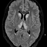
Uraemic encephalopathy with the lentiform fork sign! Results from accumulated uraemic toxins and acid – base / electrolyte derangement in the context of renal failure. There are three general (and overlapping) MRI patterns:
1⃣ Basal ganglia (most “classic”).
2⃣ Cortical - subcortical (PRES-like, more parieto-occipital).
3⃣ White-matter (centrum semiovale).

Patient with end-stage renal failure and frequent non-attendance at dialysis presenting with confusion and seizures. Bilateral swollen, T2 hyperintense lentiform nuclei with a brighter rim tracing the internal and external capsules.
What is the sign and the diagnosis?


This is NMOSD!
⭐️ NMOSD is an autoimmune astrocytopathy. AQP4-IgG targets aquaporin-4 on astrocyte endfeet; hence, lesion distribution tracks AQP4 density.
⭐️ It is rare (0.5 – 10 / 100,000), with a marked female predominance (as high as 9:1). Peak incidence is in middle age, not the classic young-adult MS demographic. Coexistent autoimmunity (e.g., SLE, Sjögren's, myasthenia) is common.
⭐️ When presenting with optic neuritis, long-segment posterior disease involving the chiasm and tracts is typical.
⭐️ Radiology "sweet spots":
- Periependymal surfaces, hypothalamic-diencephalic lesions and involvement of the area postrema in particular.
- Corticospinal tracts.
⭐️ On your axial T2, look for dot-like foci of hyperintensity in areas of spinal cord involvement, so-called "bright spotty lesions".
⭐️ Some MS treatments (e.g., interferon-β, natalizumab, fingolimod) can worsen NMOSD, so differentiation is important. B-cell depletion and complement / IL-6 blockade are the NMOSD path, not interferon or natalizumab.
This is a 40 year-old female presenting with a severe, rapidly progressive myelopathy and intractable nausea, vomiting, and hiccups. On MR there's FLAIR hyperintensity in the area postrema and a longitudinally extensive cervical spinal cord lesion.
What's the diagnosis and required test? This disease also commonly presents with optic neuritis; what's the typical radiological pattern of optic pathway involvement?


This is a 40 year-old female presenting with a severe, rapidly progressive myelopathy and intractable nausea, vomiting, and hiccups. On MR there's FLAIR hyperintensity in the area postrema and a longitudinally extensive cervical spinal cord lesion.
What's the diagnosis and required test? This disease also commonly presents with optic neuritis; what's the typical radiological pattern of optic pathway involvement?
Spot on, everyone! This is typical Wernicke's. Traditional teaching is to give thiamine before glucose. Glucose is converted to pyruvate in glycolysis; thiamine is the "key" that allows pyruvate to be processed in the Krebs cycle. Without adequate thiamine, pyruvate accumulates and is converted to lactate, so the administered glucose produces little useful energy and worsens cellular stress. In reality, profound hypoglycaemia is extremely damaging and should be corrected urgently.
A 52 year-old with a history of bariatric surgery presents with confusion, nystagmus, and ataxia. On MR we see symmetrical T2/FLAIR hyperintensity in the caudate nuclei, medial thalami, and periaqueductal grey matter. Tiny haemorrhages are scattered near the ependymal surfaces of the medial thalami.
What's the diagnosis? What's treatment needs to be given first?


A 52 year-old with a history of bariatric surgery presents with confusion, nystagmus, and ataxia. On MR we see symmetrical T2/FLAIR hyperintensity in the caudate nuclei, medial thalami, and periaqueductal grey matter. Tiny haemorrhages are scattered near the ependymal surfaces of the medial thalami.
What's the diagnosis? What's treatment needs to be given first?
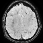
ICU MRI. The T2-weighted images are normal but the SWI looks like iron filings scattered over a magnet. Before you think amyloid angiopathy or diffuse axonal injury, pause...
These are critical illness–associated microbleeds.
Typical pattern:
✅ Juxtacortical white matter and corpus callosum.
🚫 Cortex, periventricular white matter, basal ganglia, or thalami.
❓ Internal capsules and cerebellum.
This appears up after respiratory failure, ARDS, prolonged ventilation, or ECMO, not after a fall, and not only in the elderly. Fat embolism is the main radiological mimic but comes with a "starry sky" DWI pattern.


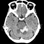
Acute non-traumatic cerebellar haemorrhage in a young patient. Nothing seen on the CT angiogram or the post-contrast MRI, but look at the ASL...
There's high signal in the occipital sinus; this is not normal. If veins light up on ASL, that means labelled arterial blood got there too fast, which usually means it skipped the capillary bed. This implies arteriovenous shunting!
Lo and behold, the DSA shows a left perimesencephalic arteriovenous malformation supplied by basilar perforators and branches from the left superior cerebellar artery, draining into the left basal vein, vein of Galen, and straight sinus.


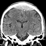
Extradural haemorrhage never crosses sutures... except when it does!
This is a typical venous extradural haemorrhage at the vertex, crossing the sagittal suture and inferiorly displacing the superior sagittal sinus and nearby cortical veins. Look carefully for active venous haemorrhage on the CTV!
You can also see venous extradural haemorrhage along the anterior margin of the middle cranial fossa and in the posterior fossa.


Interesting commentary in The Lancet. Where is the line between neurology and psychiatry? Into which category does dementia fall? Who is best placed to manage functional neurological disorders? Is there an argument to fold the DSM into the ICD? Good questions, relevant to those in the field.
thelancet.com/journals/lance…
Key reading for neuroradiologists. Demonstrates the link between quality of resection in grade 2 astrocytomas and oligodendrogliomas and survival. The benefit is greater for astrocytomas than oligodendrogliomas and becomes apparent earlier. The authors produced a four-tier system for extent of resection based on residual T2-FLAIR volume:
Class 1⃣: supramaximal (no residual tumour and resection of non-affected structures beyond imaging borders).
Class 2⃣: maximal (0 - 5 cm³ residual).
Class 3⃣: submaximal (5 - 25 cm³ residual).
Class 4⃣: minimal (> 25 cm³ residual and/or residual contrast-enhancing tumour).
thelancet.com/journals/lanon…

Jack Garnham retweeted

It’s July and that means this #tipsfornewdocs should be posted in all the work rooms

This patient presented with left-sided internuclear ophthalmoplegia (INO). How can we explain this?
👁️👁️ How normal horizontal eye movements work:
⭐️ To look to the right, the brain (mainly from the left frontal eye field) activates the right horizontal gaze center - in the paramedian pontine reticular formation (PPRF) - located in the pons.
⭐️ The right PPRF stimulates the adjacent right abducens nucleus (CN VI).
⭐️ The right abducens nucleus does two things simultaneously:
1⃣ It sends signals to the right lateral rectus muscle → right eye abducts.
2⃣ It sends interneurons that cross the midline and ascend in the left medial longitudinal fasciculus (MLF) to reach the left oculomotor nucleus (CN III, medial rectus subnucleus) in the midbrain.
⭐️ This activates the left medial rectus muscle → left eye adducts.
⭐️ This ensures both eyes move together smoothly to the right!
👁️❌👁️ What happens with a left MLF lesion?
⭐️ A lesion in the left MLF (whether in the pons or midbrain) interrupts the pathway.
⭐️When looking to the right, the right eye abducts normally (intact right CN VI), BUT...
❌The signal fails to reach the left CN III medial rectus subnucleus through the damaged left MLF.
❌Result: impaired adduction of the left eye (it lags behind or stays near the midline).
The left MLF is taken out by this infarct!


Subcortical FLAIR hypointensity is an interesting and under-recognised finding. Often seen underlying a subdural collection. Most commonly associated with seizures (including status epilepticus or subclinical seizures). Typically reversible.

Ectopic posterior pituitary. Prevalence of 1 in 4,000 - 20,000 births. Look for an absent, truncated, or thin infundibulum, a small adenohypophysis, and an ectopic extrasellar T1 hyperintense neurohypophysis, typically located along the truncated stalk or median eminence of the tuber cinereum.
Continue the search elsewhere for a Chiari 1 malformation (20%), olfactory hypoplasia, migrational anomalies, an absent septum pellucidum, ocular dysgenesis, hypoplastic optic nerves, etc.
Consider EPP in a hypoglycaemic neonate! That was the presentation in this case.

PMD, the prototypical hypomyelinating leukodystrophy. X-linked, caused by mutations in the PLP1 gene. Primarily affects males and presents as a spectrum, with severe connatal forms (neonatal onset, poor prognosis) to classic forms (infantile onset, slower progression).
White matter remains T1 hypointense or isointense to mildly hyperintense but is markedly T2 hyperintense. Myelin may be preserved in some structures (e.g., the posterior limb of the internal capsule, optic radiations, and proximal corticospinal tracts). Hypomyelination sometimes requires repeat imaging (six months, with one scan after 1 or 2 years of age) to differentiate it from delayed myelination.


A positive brain death CTA. No filling of the cortical (M4) branches of either middle cerebral artery and no filling of either internal cerebral vein. The opacification of the bilateral M1 and A1 segments is termed "stasis filling" and does not reflect effective parenchymal perfusion.
Read the BSNR guidelines here: doi.org/10.1016/j.crad.2022.….


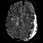
Long-segment anterior optic neuritis and multifocal cortical swelling and FLAIR hyperintensity in a four-year-old. This was MOGAD. It presents more typically with an ADEM phenotype (60 - 70%) but we also see cortical encephalitis and sometimes a leukodystrophy-like pattern. We find MOG positivity in one-third of children presenting with an initial acute demyelinating episode. In ADEM phenotypes, this rises to 40 – 60%.


