

Digital Pathologist 🔬 Medical Director 🔬 #GIpath fellowship-trained 🔬 Independent contractor 🔬 Available for remote pathology work
Detroit, MI
Joined March 2019
- Tweets2.1K
- Following1.6K
- Followers3.5K
- Likes8K




Pinned Tweet
Welcome to my first #PathTweetorial!
HOW TO PREPARE FOR PATHOLOGY BOARDS
Now that I've finished AP and CP #PathBoards, I would like to share some tips that might make it easier for those who will be taking it in subsequent years!
My tips will cover the following:
#yAdvice

In case anyone is wondering where I’ve gone… the answer is LinkedIn!
Check out my profile there to connect and see what I’m up to!
linkedin.com/in/ymdphd
4
393

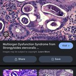
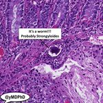
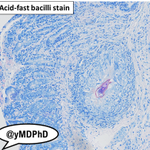
🚨Cool case🚨Duodenal biopsy with a huge epithelioid granuloma in the submucosa. Otherwise looks pretty unremarkable. Ordered deeper levels and routine bug stains.

I inspect the other levels more carefully, and then I start noticing something lurking in the background...


What say you about this serrated polyp that's too blue? If you've heard of MLH-1, that's a big clue. Answer below...

Answer: This is a sessile serrated lesion with low-grade dysplasia, confirmed with loss of nuclear MLH-1 staining in the dysplastic crypts.
I look at striking images like this every day while diagnosing patients via #DigitalPathology
Reach out if you'd like to connect!
Here's a stomach biopsy with a normal appearance on one side, and an abnormal appearance on the other. Which is which? (Answer below)

Answer: The left side is abnormal, exhibiting a reactive gastropathy pattern (i.e. chemical gastropathy). Notice the loss of clear cytoplasmic mucin of surface epithelial cells. This injury is commonly due to medications (NSAIDs) and can be associated with peptic ulcer disease.
I look at striking images like this every day while diagnosing patients via #DigitalPathology
Reach out if you want to know more about my digital diagnostic services!
Connect with me on LinkedIn linkedin.com/in/ymdphd/
1
7
769
There are too many nuclei in the colonic crypts of this patient with chronic diarrhea. If you can recognize the cells, you'll know the diagnosis (answer below!).

Answer: There are too many lymphocytes in this beautiful example of lymphocytic colitis.
I look at striking images like this all day while diagnosing patients via #DigitalPathology
Reach out if you want to know more about my digital diagnostic services!
25
1,242

Should we consider a “non disparagement” clauses in employment agreements to be a red flag?
On the flip side, would the absence of one give you a boost of confidence that a potential employer treats their people well?
2
3
626
I think there is a worsening pathologist shortage.
I’m hearing about burnout and early retirements in smaller groups.
I get pinged daily with new vacancies and locum positions all over the map.
Are our folks in larger places feeling it too?
3
20
1,314
Youssef Farhat, MD/PhD retweeted

We are back with "Meet Our Experts" It's time to get to know our Diagnexia experts a little better!
This week we are excited to introduce Dr. Youssef Farhat (@yMDPhD), a renowned expert in GI, Liver and Pancreatic Pathology.
Find out more: news.diagnexia.com/3GuWk43
#PathTwitter

Curious finding in an anal polyp on top of a hypertrophied papilla… what is all that pink stuff in the superficial squamous cells? Looks reactive, but the appearance is so striking I feel like it deserves a name…


For those who are scratching your head like I was, I think we have an answer:
Toto bodies!
Do you agree? Reference: ncbi.nlm.nih.gov/pmc/article…

2
842