





DrPrerna retweeted

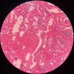
Case of Microglandular Adenosis (MGA) 🔬
An infiltrative low-grade epithelial proliferation with central eosinophilic material, devoid of myoepithelial cell layer, MGA can mimic invasive carcinoma—however, negative ER should raise suspicion🕵️♂️.
#PathX #PathTwitter #breastpath


DrPrerna retweeted

Just a simple advice on a common issue in Salivary Gland FNA - “Pleomorphic Adenoma enjoys CHAOS - disregard pleomorphism, hypercellularity, and high power atypia - PA loves to appear malignant”.
DrPrerna retweeted

You may just want to call it a “wart” or “ squamous papilloma “ in genital or oral areas and let go, No, it is verruciform xanthoma (疣状黄瘤)1) oral or genital location 2) warty like surface. 3) bright pink parakeratosis with neuts 4) foamy cells in papillary dermis.#Dermpath


DrPrerna retweeted
"Eyeballs" - Naked nuclei with macronucleoli + Granular smear background + Liver lesion = Hepatocellular Carcinoma.


DrPrerna retweeted
Intranuclear inclusions are created by redundancy of nuclear membrane leading to invagination/protrusion of the cell cytoplasm through the nucleus. This INCI from a cystic subtype of Papillary Thyroid CA proves that fact and even has the cytoplasmic vacuoles seen within it. (FNA)


DrPrerna retweeted

Sessile serrated polyp, sessile serrated adenoma, sessile serrated lesion, Oh whatever! #Gipath
DrPrerna retweeted

MYXOID LIPOSARCOMA. NGS: FUS-DDIT3. NB: spindle-stellate cells set within abundant myxoid stroma; pools of extracellular mucin with a so-called 'pulmonary oedema' pattern; delicate branching vasculature; scattered small lipoblasts.


DrPrerna retweeted

Before we (hopefully) get to turn off your brains for the holiday, here's a quick lesson!
Characteristic vascular patterns in #bstpath:
"Chicken wire" ➡️ Myxoid liposarcoma*
"Curvilinear" ➡️ Myxofibrosarcoma
(*I agree w/ @JMGardnerMD that they look more like chicken feet)


DrPrerna retweeted
Gynecologic PEComa (Malignant):
🔬 Spindle + epithelioid cells
🔬 Nests, sheets, fascicles
🔬 Atypia: low to anaplastic
🔬 ≥1 pattern of invasion
📝 Grading [see table]
🎨 Melanocytic + smooth musc IHC
🧬 TSC1/2 mutn, TFE3 fusion
#gynpath #bstpath #pathtweetorial @EmoryPathology


DrPrerna retweeted

PEComa of uterus – diffuse, nested, or corded growth of epithelioid/spindle cells with prominent vascularity and perivascular radial arrangement of tumor cells. Co-expression of smooth muscle and melanocytic markers on IHC. Associated with TSC1/2, TFE3 mutations. #GYNPath

DrPrerna retweeted

Squamous metaplasia of cervix
1. Epithelialisation: direct in growth from portion cx
2. Metaplasia: subcolumnar reserve cells... Maturation into immature... mature squamous cells
Etiology :Irritation, trauma, infection
Dd: SIL (atypia+, endocervical cells - )
#gupath #pathology


DrPrerna retweeted
If you think THYROID Cytology is easy ...
Left - Medullary Carcinoma (MTC)
Right - Hurthle Cell Neoplasm (HCN) - Carcinoma on follow-up.
INCIs maybe helpful for an MTC dx, as they are EXCEEDINGLY rare in HCNs (In fact, for many years I believed that you never see them in HCNs.


DrPrerna retweeted
AMAZING aspiration done by our radiologist from a 7mm thyroid nodule. No, it’s not a lymphoma or intrathyroidal thymic lesion. Higher power clearly shows plasmacytoid cells consistent with “Medullary Thyroid Carcinoma”. Perhaps, the most cellular MTC that I’ve ever seen on FNA.


DrPrerna retweeted
"Nothing is outright specific in Cytomorphology" - Papillary thyroid CA (PTC) on FNA displaying the impressive so-called "Intracytoplasmic Colloid Inclusions" (ICI). ICI has been considered a marker for "Hurthle Cell Neoplasm" but I have seen them numerous times in PTCs.

DrPrerna retweeted

T is for tangles "lymphoid tangles"=clusters/aggregates of crushed #lymphocyte nuclei as frequently seen in lymphoid lesions but a useful feature for #lymphocytic thyroiditis and #Warthin tumour as in this case 👇#CytoABCs #pathology trainees #cytopath #citologia #PAAF #FNA


#CytoABCs!
T is for Troll hair, named for frizzy hair on the kids' toys, describing the fibrillary, bright magenta metachromatic stroma seen in pleomorphic adenomas on Romanowsky stains (like DQ seen here). On Pap stain (this one also has tyrosine crystals!) the stroma is pale.


DrPrerna retweeted
"The Scorpion and the Ants" - Thyroid FNA can be extremely challenging when facing the dilemma of "Medullary CA versus Hurthle Cell Neo". This case, despite hypercellularity, single cells and lack of nucleolar prominence, turned out to be "Hurthle Cell CA" on resection.


